Bilateral spontaneous rectus sheath haematoma complicating dengue haemorrhagic fever: a case report.
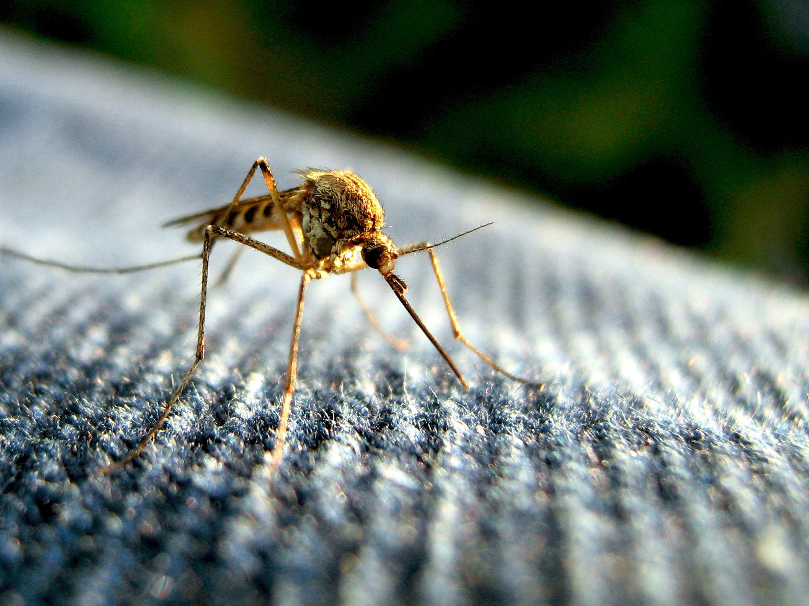
Dengue is the most important mosquito-borne viral disease affecting humans and can be caused by any 1 of 4 dengue viruses (DENV-1 to DENV-4), transmitted by Aedes mosquitoes [1]. Currently, about 100 million clinically apparent cases are estimated to occur each year resulting in approximately 20000 deaths [2].
Rectus sheath haematoma (RSH) is an uncommon entity which, when occurring without any antecedent history of trauma is referred to as spontaneous RSH. It usually occurs in the lower quadrants of the abdominal wall and almost never crosses the midline. Anticoagulation therapy, blood dyscrasias, pregnancy, blunt trauma, abdominal straining, obesity and coughing are some common risk factors for RSH [3]. We present the first case of spontaneous and bilateral rectus sheath haematoma complicating dengue haemorrhagic fever (DHF) in an elderly male at the beginning of the defervescence phase of his viral illness who was managed conservatively.
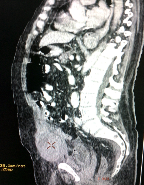
Case report: A 72-year elderly male presented in October 2013 with the complaint of fever and headache for four days. He also had low back pain with generalised myalgias for last 2 days. There was no history of vomiting, cough, convulsions or any bowel or bladder symptoms. His illness overlapped a recent dengue outbreak that occurred in North India from August 2013 to November 2013. On examination, he was noted to have an oral temperature of 38.2 oC, pulse rate of 82 beats per minute and blood pressure of 138/90 mmHg. He had no jaundice, neck stiffness or any rash. The rest of his general and systemic examination was unremarkable. Laboratory investigations revealed haemoglobin of 127 g/L, haematocrit – 46.6 %, total leucocyte count of 5.6×10⁹/L (differential count ; normal), platelet count of 60×109/L and ESR of 40 mm at the end of 1st hour. Serum levels of bilirubin, aspartate transaminase, alanine transaminase and alkaline phosphatase were 20.52µmol/L, 86 IU/L, 48 IU/L and 96 IU/L respectively. He tested negative for Malaria (Optimal –IT), HIV-1 and 2 (ELISA), HBsAg and anti- HCV antibody. The early NS1 antigen detection test and IgM antibody assay (ELISA) for dengue fever was positive. Abdominal ultrasonography was unremarkable. With a high suspicion of dengue fever, the patient was put on conservative management. His fever and back pain subsides after four days and blood count profile done one week later revealed haemoglobin of 120 g/L, haematocrit – 42.6 % and a platelet count of 100×109/L. However a day after, prior to the discharge, he complained of gradually worsening lower abdominal pain and difficult micturition. He was afebrile and his blood pressure was 110/80 mmHg. Abdominal examination revealed a tender, firm mass in the infra-umbilical area. His haemoglobin level when repeated, revealed a fall from 120 g/L to 92 g/L and a haematocrit of 32% with platelet count of 90×109/L. Abdominal pain along with an acute drop in the haemoglobin level and without any evidence of overt blood loss raised the suspicion of intra-abdominal bleeding. Ultrasound of the lower abdomen revealed a large collection in the anterior wall (Figure 1a) and contrast-enhanced abdominal CT scan confirmed a bilateral, spindle-shaped, thickened rectus sheath consistent with RSH (Figure 2 and 3). He had no history of any bleeding disorder, trauma or exposure to anti-coagulant drugs and his coagulation profile was normal. The temporal relation of the events, corroborative drop in haemoglobin, positive dengue serology and thrombocytopenia were all consistent with RSH complicating dengue haemorrhagic fever. Over the next eight hours, he developed diffuse ecchymotic area over his lower abdomen simulating Turner’s and Cullen’s sign (figure 1b). He was kept under close follow up and received three units of blood transfusion while angiographic embolization was planned as a backup. The patient was fairly stable as there was no further increase in the size of RCH. He recovered uneventfully and was discharged one week later.
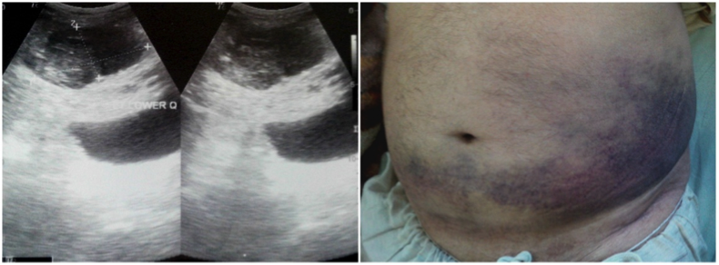
Figure 2: Computed tomography (CT) image demonstrating a bilateral rectus sheath haematoma.
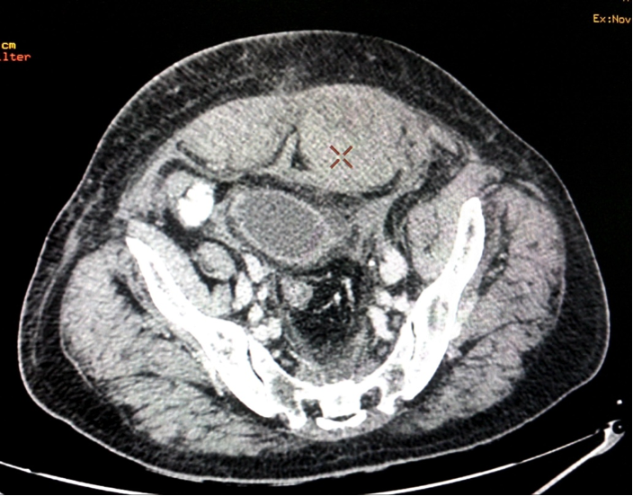
Figure 3: CT scan (sagittal section) showing a large rectus sheath haematoma.
Discussion:
Haemorrhagic manifestations in patients with dengue fever are not uncommon and range from petechiae and purpura to severe gastrointestinal haemorrhage and shock. The pathophysiological basis of haemorrhage in dengue infections remains poorly understood, despite its increasing global burden. Bleeding in patients with dengue infections may result from a combination of thrombocytopenia, low circulating levels of proteins C, S, and antithrombin III, dysfunctional surviving platelets, and increased fibrinolysis [4].The critical stage of DHF is the time of defervescence when fever abates and signs of circulatory failure or haemorrhagic manifestations occur, possibly going unnoticed [5].
The clinical peculiarities of dengue fever in the elderly are less clearly known. Elderly hosts are subjected to higher risk of infection-related mortality and morbidity associated with dengue fever and complications like shock, concurrent bacteremia, coagulation abnormalities, gastrointestinal bleeding and acute renal failure are not uncommon [6]. Prothrombin time has also been found to be prolonged exclusively in the elderly dengue patients increasing the risk of bleeding [4].
RSH of the anterior abdominal wall is caused by the rupture of superior or inferior epigastric artery, their branches or a tear of the rectus muscle. It is usually frequent and self-limiting in elderly females, however, it can cause an expanding haematoma turning into a haemoperitoneum [3]. For those receiving anticoagulation therapy, the mortality rate has been reported to be as high as 25% [7]. It usually occurs unilaterally, but a few rare cases of bilateral haematoma have been reported. [8] A thorough literature search revealed only two cases of unilateral spontaneous RSH attributing to DHF [9-10]. Our case is probably the first case of bilateral spontaneous RSH complicating DHF in an elderly male at the beginning of the defervescence phase of his viral illness who was promptly managed. He also developed ecchymoses in the flanks and periumbilical area due to the dissolution of the haematoma in the adjoining planes simulating Gray Turner’s and Cullen’s signs respectively, rare late signs. RSH can pose a diagnostic difficulty and CT scan is the diagnostic modality of choice, although ultrasonography is a suitable alternative [10]. Conservative treatment including intravenous hydration, blood transfusion and strict monitoring are appropriate in most of the settings with abdominal wall haematomas. Surgical intervention or transcatheter arterial embolization is recommended only when conservative management fails [7].
Key points/conclusion.
RSH is a great masquerader and can mimic most other abdominal emergencies. Unnoticed and fatal complications can occur during the dengue viral illness in the elderly and hence a high index of “suspicion and supervision” should be maintained during its defervescence phase. Prompt diagnosis and astute judgment in the context of clinical awareness usually suffice and negate the need of any surgical intervention which can further complicate the outcome in the elderly population.
Bibliography: 1 Simmons CP, Farrar JJ, Nguyen VV, Wills B Dengue. N Engl J Med 2012;366:1423-32.
2 Bhatt S, Gething PW, Brady OJ, Messina JP, Farlow AW, Moyes CL,et al. The global distribution and burden of dengue. Nature 2013;496:504
3 Cherry WB, Mueller PS. Rectus sheath hematoma, review of 126 cases at a single institution. Medicine (Baltimore) 2006; 85 :105-10.
4 Wills BA, Oragui EE, Stephens AC, Daramola OA, Dung NM, Loan HT, et al. Coagulation abnormalities in dengue hemorrhagic fever: serial investigations in 167 Vietnamese children with dengue shock syndrome. Clin Infect Dis 2002;35:277-85.
5 Martina BE, Koraka P, Osterhaus AD.Dengue virus pathogenesis: an integrated view. Clin Microbiol Rev 2009 ;22 :564-81
6 Lee IK, Liu JW, Yang KD. Clinical and Laboratory characteristics and risk factor for fatality in elderly patients with dengue haemorrhagic fever. Am J Trop Med Hyg 2008; 79 : 149–53.
7 Yamada Y, Ogawa K, Shiomi E and Hayashi T. Images in cardiovascular medicine. Bilateral rectus sheath hematoma developing during anticoagulant therapy. Circulation 2010;121:1778-79
8 Auten JD, Schofer JM, Banks SL, Rooney TB. Exercise-induced bilateral rectus sheath hematomas presenting as acute abdominal pain with scrotal swelling and pressure: case report and review. J Emerg Med 2010 ; 38:e9-12.
9 Tong P, Yeoh CJ, Yong EL Abdominal mass and a forgotten haemorrhagic fever. Lancet 2010; 376: 140
10 Ganeshwaran Y, Seneviratne SM, Jayamaha R, De Silva AP, Balasuriya WK. Dengue fever associated with a haematoma of the rectus abdominis muscle. Ceylon Med J 2001;46:105-6.
- Log in to post comments
